ELBOW

CONDITIONS

  • ELBOW DISLOCATION
  • GOLFER’S ELBOW
  • TENNIS ELBOW
ELBOW DISLOCATION

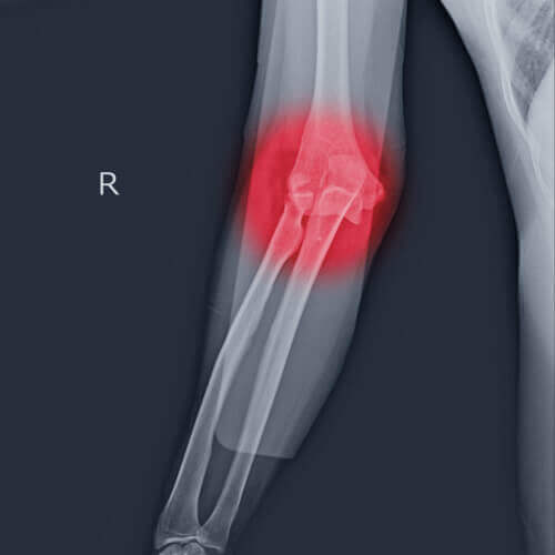
The elbow is a hinge joint made up of 3 bones – humerus, radius and ulna. The bones are held together by ligaments to provide stability to the joint. Muscles and tendons move the bones around each other and help in performing various activities. Elbow dislocation occurs when the bones that make up the joint are forced out of alignment.

Elbow dislocations usually occur when a person falls onto an outstretched hand. Elbow dislocations can also occur from any traumatic injury such as motor vehicle accidents. When the elbow is dislocated you may have severe pain, swelling, and lack of ability to bend your arm. Sometimes you cannot feel your hand, or may have no pulse in your wrist because arteries and nerves run along your elbow may be injured.

To diagnose elbow dislocation your doctor will examine your arm. Your doctor will check the pulses at the wrist and will evaluate the circulation to the arm. An X-ray is necessary to determine if there is a break in the bone. An arteriogram, an x-ray of your artery can be helpful to know if the artery is injured.

An elbow dislocation is a serious injury and therefore requires immediate medical attention. At home, you may apply an ice pack to the elbow to ease pain and swelling. However it is important to see your doctor for help. You can also check if the arteries and nerves are injured or remain intact. You can feel your pulse by pressing tips of your fingers at the base of your wrist. They should turn white or blanch and a pink color should come back in 3 seconds. To check for nerves, first bend your wrist up and move your fingers apart and then touch your thumb to your little finger. You can also check for numbness all over your hand and arm. If you have problem with any of these tests you need to see your doctor right away.

You doctor will put your dislocated elbow back in place by pulling down your wrist and levering your elbow. This procedure is known as reduction. As it is a painful procedure you may be given medications to relieve your pain before the procedure. After the reduction you may have to wear a splint to immobilize your arm at the elbow. After few days you may also need to do gentle motion exercises to improve the range of motion and strength.

Elbow dislocations may be prevented if you avoid falling on outstretched arm or avoid situations that may cause falls such as walking at night or walking on slippery floors.

GOLFER’S ELBOW

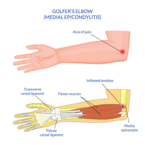
Golfer’s elbow, also called Medial Epicondylitis, is a painful condition occurring from repeated muscle contractions in the forearm that leads to inflammation and microtears in the tendons that attach to the medial epicondyle. The medial epicondyle is the bony prominence that is felt on the inside of the elbow.

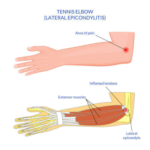
Golfer’s elbow and Tennis Elbow are similar except that Golfer’s elbow occurs on the inside of the elbow and Tennis Elbow occurs on the outside of the elbow. Both conditions are a type of Tendonitis which literally means “inflammation of the tendons”.

Signs and symptoms

Signs and symptoms of Golfer’s Elbow can include the following:

  • Elbow pain that appears suddenly or gradually
  • Achy pain to the inner side of the elbow during activity
  • Elbow stiffness with decreased range of motion
  • Pain may radiate to the inner forearm, hand or wrist
  • Weakened grip
  • Pain worsens with gripping objects
  • Pain is exacerbated in the elbow when the wrist is flexed or bent forward toward the forearm

Causes

Golfer’s Elbow is usually caused by overuse of the forearm muscles and tendons that control wrist and finger movement but may also be caused by direct trauma such as with a fall, car accident, or work injury.

Golfer’s elbow is commonly seen in golfer’s, hence the name, especially when poor technique or unsuitable equipment is used when hitting the ball. Other common causes include any activity that requires repetitive motion of the forearm such as: painting, hammering, typing, raking, pitching sports, gardening, shoveling, fencing, and playing golf.

Diagnosis

Golfer’s Elbow should be evaluated by an orthopaedic specialist for proper diagnosis and treatment.

  • Medical History
  • Physical Examination
  • Your physician may order an x-ray to rule out a fracture or arthritis as the cause of your pain.
  • Occasionally, if the diagnosis is unclear, your physician may order further tests to confirm golfer’s elbow such as MRI, ultrasonography, and injection test

Conservative Treatment Options

Your physician will recommend conservative treatment options to treat the symptoms associated with Golfer’s Elbow. These may include the following:

  • Activity Restrictions: Limit use and rest the arm from activities that worsen symptoms
  • Orthotics: Splints or braces may be ordered to decrease stress on the injured tissues
  • Ice: Ice packs applied to the injury will help diminish swelling and pain. Ice should be applied over a towel to the affected area for 20 minutes four times a day for a couple days. Never place ice directly over the skin
  • Medications: Anti-inflammatory medications and/or steroid injections may be ordered to treat the pain and swelling
  • Occupational Therapy: OT may be ordered for strengthening and stretching exercises to the forearm once your symptoms have decreased
  • Pulsed Ultrasound: A non-invasive treatment used by therapists to break up scar tissue and increase blood flow to the injured tendons to promote healing
  • Professional instruction: Consulting with a sports professional to assess and instruct in proper swing technique and appropriate equipment may be recommended to prevent recurrence

Surgery

If conservative treatment options fail to resolve the condition and symptoms persist for 6 -12 months, your surgeon may recommend surgery to treat Golfers Elbow. The goal of surgery to treat Golfers Elbow is to remove the diseased tissue around the inner elbow, improve blood supply to the area to promote healing, and alleviate the patient’s symptoms.

TENNIS ELBOW

Tennis elbow is the common name used for the elbow condition called lateral epicondylitis. It is an overuse injury that causes inflammation of the tendons that attach to the bony prominence on the outside of the elbow (lateral epicondyle).It is a painful condition occurring from repeated muscle contractions at the forearm that leads to inflammation and micro tears in the tendons that attach to the lateral epicondyle. The condition is more common in sports activities such as tennis, painting, hammering, typing, gardening and playing musical instruments. Patients with tennis elbow experience elbow pain or burning that gradually worsens and a weakened grip

Your doctor will evaluate tennis elbow by reviewing your medical history, performing a thorough physical examination and ordering X-rays, MRI or electromyogram (EMG) to detect any nerve compression.

Your doctor will first recommend conservative treatment options to treat the tennis elbow symptoms. These may include:

  • Limit use and rest the arm from activities that worsen symptoms.
  • Splints or braces may be ordered to decrease stress on the injured tissues.
  • Apply ice packs on the elbow to reduce swelling.
  • Avoid activities that bring on the symptoms and increase stress on the tendons.
  • Anti-inflammatory medications and/or steroid injections may be ordered to treat pain and swelling.
  • Physical therapy may be ordered for strengthening and stretching exercises to the forearm once your symptoms have decreased.
  • Pulsed ultrasound may be utilized to increase blood flow and promote healing to the injured tendons.

If conservative treatment options fail to resolve the condition and symptoms persist for 6 -12 months, your surgeon may recommend a surgical procedure to treat tennis elbow called lateral epicondyle release surgery. Your surgeon will decide whether to perform your surgery in the traditional open manner (single large incision) or endoscopically (2 to 3 tiny incisions and the use of an endoscope –narrow lighted tube with a camera).Your surgeon will decide which options are best for you depending on your specific circumstances.

Your surgeon moves aside soft tissue in order to view the extensor tendon and its attachment on the lateral epicondyle. The surgeon then trims the tendon or releases the tendon and then reattaches it to the bone. Any scar tissue present will be removed as well as any bone spurs. After the surgery is completed, the incision(s) are closed by suturing or by tape.

Following surgery, you are referred to physical therapy to improve the range of motion and strength of your joint.


TREATMENT

  • ELBOW ARTHROSCOPY
ELBOW ARTHROSCOPY

Elbow arthroscopy, also referred to as keyhole or minimally invasive surgery, is performed through tiny incisions to evaluate and treat several elbow conditions.

The Elbow is a complex hinge joint formed by the articulation of three bones – humerus, radius and ulna. The upper arm bone or humerus connects the shoulder to the elbow forming the upper portion of the hinge joint. The lower arm consists of two bones, the radius and the ulna. These bones connect the wrist to the elbow forming the lower portion of the hinge joint.

The three joints of the elbow are

  • Ulnohumeral joint, the junction between the ulna and humerus
  • Radiohumeral joint, the junction between the radius and humerus
  • Proximal radioulnar joint, the junction between the radius and ulna

The elbow is held in place with the support of various soft tissues including:

  • Cartilage
  • Tendons
  • Ligaments
  • Muscles
  • Nerves
  • Blood vessels and
  • Bursae

Indications of elbow arthroscopy:

Elbow arthroscopy is usually recommended for the following reasons:

  • Debridement of loose bodies such as bone chips or torn cartilage tissue
  • Removal of scar tissue
  • Removal of bone spurs: These are extra bony growths caused by injury or arthritis that damage the ends of bones causing pain and limited mobility.

Arthroscopy is also used for the:

Treatment of osteoarthritis, rheumatoid arthritis, and a condition called osteochondritis dissecans where loose fragments of cartilage and bone are in the joint space.

Evaluation and Diagnosis:

Your surgeon will review your medical history and perform a complete physical examination. Diagnostic studies may also be ordered such as X-rays, MRI or CT scan to assist in diagnosis.

Surgical Procedure

Arthroscopy is a surgical procedure in which an arthroscope, a small soft flexible tube with a light and video camera at the end, is inserted into a joint to evaluate and treat a variety of conditions.

Elbow arthroscopy is commonly performed under general anesthesia as an outpatient procedure. The patient is placed in a lateral or prone position which allows the surgeon to easily adjust the arthroscope and have a clear view of the inside of the elbow.

Several tiny incisions are made to insert the arthroscope and small surgical instruments into the joint. To enhance the clarity of the elbow structures through the arthroscope, your surgeon will fill the elbow joint with a sterile liquid.

The liquid flows through the arthroscope to maintain clarity and also to restrict any bleeding. The camera attached to the arthroscope displays the internal structures of the elbow on the monitor and helps your surgeon to evaluate the joint and direct the surgical instruments to fix the problem.

At the end of the procedure, the surgical incisions are closed by sutures, and a soft sterile dressing is applied. Your surgeon will place a cast or a splint to restrict the movement of the elbow.

The advantages of arthroscopy compared to traditional open elbow surgery include:

  • Smaller incisions
  • Minimal soft tissue trauma
  • Less post-operative pain
  • Faster healing time
  • Lower infection rate

Post-operative care:

The post-surgical instructions include:

  • Make sure to get adequate rest.
  • Raise your elbow on pillows above the level of the heart to help reduce swelling.
  • Keep the incision area clean and dry.
  • A compressive stocking may be applied from the armpit to the hand once the dressing is removed to decrease pain and increase range of motion.
  • Your doctor will prescribe pain medications to keep you comfortable.
  • Physical therapy will be ordered to restore normal elbow strength.
  • Eating a healthy diet and not smoking will promote healing.

Complications:

The possible complications following elbow arthroscopy include infection, bleeding, and damage to nerves or blood vessels.

OUR TEAM

Dr. Christopher Butcher

DR. CHRISTOPHER BUTCHER
Consultant Orthopaedic and Knee Surgeon

Dr Les Cannon trauma surgeon

PROF. DR. LESLIE CANNON
Consultant Orthopaedic and Trauma Surgeon

Dr. Lucia Heras - Hand Surgeon

DR. LUCIA HERAS
Consultant Orthopedic & Hand Surgeon

Neda Ali Physiotherapist

NEDA MOHAMMAD ALI

Physiotherapist, Musculoskeletal Specialist
shreya physiotherapist

SHREYA SINGH

Physiotherapist, Musculoskeletal Specialist
rathish physiotherapist

RATHISH MANICKAM

Musculoskeletal Physiotherapist and Specialist in Manual Therapy
Lubnah Darawsheh

LUBNAH DARAWSHEH

Physiotherapist
Ahmed Elhamedy

AHMED ALHAMEDY

Physiotherapist , Musculoskeletal Specialist and Sports Rehabilitation
sofia turner

SOFIA TURNER

Physiotherapist


BOOK AN APPOINTMENTREVIEW US ON GOOGLE

Book an Appointment


    Your Name (required)

    Your Email (required)

    Mobile No. (required)

    Select Appointment Date

    Copyright © Harley Street Medical Centre 2023 | MOH : EXYMXHJS-120723 15/07/2024

    CALL
    02 613 3999
    ONLINE
    APPOINTMENT
    BOOK AN APPOINTMENT
    Scan the code